Understanding Macular Degeneration
What it is, what you can do, and how we treat it
What is macular degeneration?
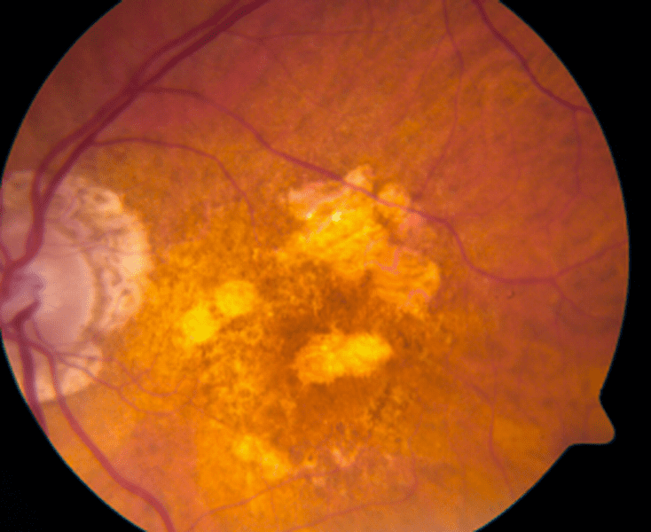
Age-related macular degeneration (AMD) is a disease of the macula. The macula is a small area in the center of the retina, the thin layer of tissue that lines the back of your eye. You use your macula for sharp, straight-ahead vision: reading, driving, seeing faces, and noticing fine details.
AMD does not cause total blindness. Your side (peripheral) vision usually stays intact. But AMD can make the center of your vision blurry, bent, or dark, and that can make everyday tasks much harder.
AMD is one of the leading causes of vision loss in adults over 50. The good news is that there is a lot you can do. Healthy habits, careful monitoring, and today's treatments can slow the disease and help protect the vision you have.
The three main forms of AMD
AMD is often described in stages. Most people start with dry AMD. Some will later develop one of the advanced forms: geographic atrophy (advanced dry) or wet AMD. It is possible to have different forms in each eye.
1. Dry AMD (early and intermediate)
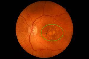
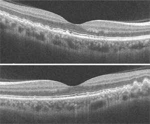
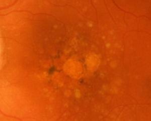
What we see in your eye: Small yellow deposits called drusen build up under the retina. Drusen are made of fats and proteins. Small drusen are common with age. Medium and large drusen, along with changes in the eye's pigment, mean a higher chance that the disease will progress. On a scan of the retina (called an OCT), drusen look like bumps under the retina's outer layer.
What you may notice:
- In the early stage, often nothing at all. This is why regular eye exams matter.
- Needing more light to read, or trouble adjusting when you walk from a bright room into a dim one.
- Mild blur in your central vision, or print that looks faded.
- Colors that seem less bright than they used to.
2. Advanced dry AMD: geographic atrophy (GA)
What we see in your eye: In geographic atrophy, patches of the retina's light-sensing cells and their support layer slowly wear away and die. On photos and scans, these patches show up as clearly outlined areas that look a bit like shapes on a map, which is how the condition got its name. GA usually grows slowly over months and years. It often starts just outside the very center of the macula and spreads.
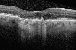
What you may notice:
- Blind spots or missing areas near the center of your vision. Letters may seem to drop out of words when you read.
- Trouble reading even with good glasses and good light.
- Trouble recognizing faces, especially from a distance.
- Vision that is often worse in dim light.
- Once the very center of the macula is affected, a dark or empty spot right where you are looking.
3. Wet AMD (neovascular AMD)
What we see in your eye: In wet AMD, abnormal blood vessels grow under or in the retina. These vessels are weak and leaky. They can leak fluid or bleed, which lifts and damages the macula. On an OCT scan, we can see the fluid as dark pockets in or under the retina. Wet AMD can cause vision loss quickly, sometimes over days or weeks. Without treatment, it can leave a scar that causes permanent central vision loss.
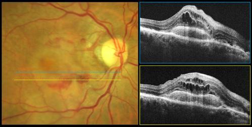
What you may notice:
- Straight lines that look wavy, bent, or broken (door frames, window blinds, lines of text).
- A sudden blurry, gray, or dark spot in the center of your vision.
- Objects that look smaller or a different size in one eye than the other.
- A fast change in vision over a few days.
|
Wet AMD is an eye emergency. If you notice new wavy lines or a new dark spot, call our office the same day. Early treatment gives you the best chance to protect your vision.
|
Quick comparison
|
|
Dry AMD
|
Geographic atrophy
|
Wet AMD
|
|
How common
|
Most people with AMD (about 8 to 9 out of 10)
|
An advanced form of dry AMD
|
Fewer people, but causes most severe and sudden vision loss
|
|
What happens
|
Drusen deposits build up under the retina
|
Patches of retinal cells wear away
|
Leaky blood vessels grow under the retina
|
|
Speed
|
Slow, over years
|
Slow, steady growth over months to years
|
Can be fast, over days to weeks
|
|
Main symptoms
|
Often none early; needs more light; mild blur
|
Blind spots near the center; trouble reading
|
Wavy lines; sudden dark or blurry spot
|
|
Treatment
|
Healthy habits, AREDS2 vitamins, Valeda light therapy (if eligible)
|
Complement inhibitor injections to slow growth
|
Anti-VEGF injections, given as a series
|
Who is at risk?
AMD is caused by a mix of genes, age, and lifestyle. Some risk factors you cannot change. Others you can, and those are where you have real power.
Risk factors you cannot change
- Risk goes up sharply after age 50 and keeps rising with each decade.
- Genetics and family history. If a parent, brother, or sister has AMD, your risk is higher. Scientists have found several genes linked to AMD, including genes that control part of the immune system called the complement system. Having these genes does not mean you will get AMD. It means healthy habits matter even more for you.
- Race and eye color. AMD is more common in white people and in people with light-colored eyes, though anyone can get it.
Risk factors you can change
- This is the single biggest risk factor you can control. See the next section.
- A diet high in leafy greens, fruits, and fish, and low in processed foods, is linked to lower risk.
- High blood pressure and heart disease. What is good for your heart and blood vessels is good for your eyes.
- Weight and activity. Being overweight and not exercising are linked to faster AMD progression. Regular walking is a great start.
Quitting smoking: the most important step
Smoking damages the blood vessels that feed the retina and floods the eye with harmful chemicals. Research shows smokers are up to four times more likely to develop AMD than people who have never smoked. Smokers also tend to get AMD earlier and see it progress faster. Smoking can make treatments less effective, too.
Here is the encouraging part: quitting helps, even if you already have AMD. People who quit have a much lower risk than those who keep smoking, and their risk keeps dropping over time. It is never too late to stop.
|
Help to quit
• Call 1-800-QUIT-NOW (1-800-784-8669) for free coaching in every state.
• Visit smokefree.gov for tools, text programs, and apps.
• Ask your primary care doctor about nicotine patches, gum, or prescription medicines. Using support roughly doubles your chances of quitting for good.
• Avoid secondhand smoke when you can.
|
Eating for your eyes
What you eat can make a difference. A large European study of nearly 5,000 adults found that people who followed a Mediterranean-style diet most closely had a 41% lower risk of developing advanced AMD than those who followed it least. No single food did the job by itself. The benefit came from the overall eating pattern.
What is the Mediterranean diet?
It is the traditional way of eating in countries around the Mediterranean Sea, such as Greece, Italy, and Spain. It is not a strict diet. It is a pattern built around plants, healthy fats, and fish.
|
Eat more of these
|
Eat less of these
|
|
• Leafy greens: spinach, kale, collards, turnip and mustard greens
• Colorful vegetables and fruits: peppers, corn, oranges, berries
• Fish and seafood, at least 2 servings a week
• Extra virgin olive oil as your main fat
• Beans, peas, and lentils
• Nuts and seeds
• Whole grains: oatmeal, brown rice, whole wheat bread
|
• Red meat and processed meats (bacon, sausage, deli meats)
• Fried foods
• White bread, white rice, and pastries
• Sugary drinks and sweets
• Butter and heavy sauces
• Highly processed snack foods
|
Fish: aim for at least 2 servings a week
Fatty fish are rich in omega-3 fats, which help keep the retina healthy. In large studies, people who ate fish regularly as part of a healthy diet were less likely to have their AMD progress. One serving is about 3 to 4 ounces, roughly the size of a deck of cards.
- Best choices: salmon, sardines, mackerel, trout, herring, and light tuna, preferably wild-caught.
- Cook it well: bake, broil, or grill. Fried fish loses much of the benefit.
- Note: Fish oil pills have not been shown to slow AMD. Eating real fish is the better choice.
Leafy greens matter too. Dark leafy greens are packed with lutein and zeaxanthin, natural pigments that collect in the macula and help shield it from damaging light. Try to eat some most days.
Eye vitamins (AREDS2 formula)
Two large studies by the National Eye Institute, called AREDS and AREDS2, tested a specific mix of vitamins and minerals. In people with intermediate AMD, or advanced AMD in one eye, the formula lowered the risk of progressing to advanced AMD by about 25%.
|
Ingredient
|
Daily amount in the AREDS2 formula
|
|
Vitamin C
|
500 mg
|
|
Vitamin E
|
400 IU
|
|
Lutein
|
10 mg
|
|
Zeaxanthin
|
2 mg
|
|
Zinc
|
80 mg
|
|
Copper
|
2 mg
|
Important things to know
- These vitamins are for people with intermediate AMD or advanced AMD in one eye. They have not been shown to help people with early AMD or to prevent AMD from starting although there are some studies that suggest they improve contrast sensitivity in early AMD. We will tell you whether they are right for you.
- If you smoke or used to smoke, choose AREDS2 or AREDS3, not the original AREDS formula. The original formula contains beta-carotene, which raised the risk of lung cancer in smokers and former smokers. AREDS2 replaced it with lutein and zeaxanthin.
- Look for "AREDS2" or “AREDS3” on the label and check that the amounts match the table above. Many "eye health" vitamins do not.
- Vitamins slow the disease. They do not cure it or restore lost vision.
- Tell your primary care doctor and pharmacist that you take them, especially if you have other health conditions.
Check your vision at home: the Amsler grid
The Amsler grid is a simple tool that helps you catch changes early, especially changes that can mean dry AMD has turned into wet AMD. Check each eye once a day. Keep the grid somewhere you will see it, such as on the refrigerator or bathroom mirror.
How to use it
- Wear your reading glasses. Use good, even lighting.
- Hold the grid about 12 to 15 inches from your face.
- Cover one eye with your hand.
- Look only at the center dot. Keep your eye on the dot the whole time.
- While looking at the dot, notice whether all the lines look straight and all the squares look the same size. Notice any wavy, blurry, or missing areas.
- Switch eyes and repeat.
- If anything looks new or different from last time, call us the same day.
|
Call us the same day if you notice:
• New wavy, bent, or broken lines on the grid or in everyday life
• A new dark, gray, blurry, or empty spot in your central vision
• A sudden drop in vision in either eye
• Objects that suddenly look a different size or shape in one eye
Southern Eye Institute: 985-872-5577
|
Treating dry AMD
For years, the only tools for dry AMD were healthy habits and vitamins. Today there are more options. Which one fits you depends on your stage of AMD and how well you see.
Valeda Light Delivery System (photobiomodulation)
Valeda is the first FDA-authorized treatment designed to improve vision in people with dry AMD. It uses gentle, low-level light at three specific colors (yellow, red, and near-infrared) to boost energy production in stressed retinal cells. This is called photobiomodulation, or PBM.
What it is like: Treatment is painless. There are no needles, no drops into the eye, and no heat. You sit at the device and look into it, much like looking into a microscope. Each treatment takes about 4 to 5 minutes per eye. Part of the time your eyes are open and you see amber and red light; part of the time your eyes are closed while a steady red light shines.
The treatment schedule
Valeda is a series of treatments, not a one-time visit. The benefit depends on completing the full schedule.
|
Step
|
What happens
|
|
One treatment series
|
9 treatments, 3 times a week, over 3 to 5 weeks
|
|
Repeat series
|
A new series about every 4 months
|
|
Full 2-year course
|
6 series, for a total of 54 treatments
|
What the research shows: the LIGHTSITE III trial
LIGHTSITE III was the main U.S. study of Valeda. It included 100 people (148 eyes) with dry AMD. Some eyes got real Valeda treatments and some got a "sham" (placebo) treatment that looked similar but did delivered 1/10th the strength of the therapeutic light. Patients were treated every 4 months for 2 years.
- Better vision on the eye chart. On average, Valeda-treated eyes gained about 6 letters on the eye chart, which is a little more than one full line. Eyes in the sham group stayed about the same, changing by about 1 letter.
- Many patients improved. About 6 in 10 treated eyes gained at least 5 letters (one line). About 1 in 5 gained 10 letters (two lines) or more.
- Fewer cases of geographic atrophy. Over 2 years, new geographic atrophy developed in about 7% of treated eyes, compared with 24% of sham eyes.
- Side effects were similar in the treated and sham groups. No serious side effects were linked to the light treatment.
|
Setting realistic expectations. Results vary from person to person. Some patients see a clear gain in vision. For others, the goal is to keep vision stable. The average gain in the study was modest, about one line on the eye chart, and the benefit appears to depend on staying on the repeat schedule. Valeda does not treat wet AMD. Overall, 85% of the treated patients either improved or remained the same which differs significantly from the vast majority of AMD patients who slowly worsen over time
|
Who may be a good candidate for Valeda?
- People with dry AMD with medium or large drusen, or geographic atrophy that does not involve the very center of the macula
- Vision between 20/32 and 20/70 with glasses
Valeda is not right for people who:
- Have wet AMD, or geographic atrophy in the very center of the macula
- Are sensitive to light, or have seizures or migraines triggered by light
- Have used certain light-sensitizing medicines or treatments in the past 30 days (please bring a list of all your medicines)
Complement inhibitor injections for geographic atrophy
The complement system is part of your immune system. In people with geographic atrophy, it appears to be overactive in the retina and helps drive the damage. Two FDA-approved medicines calm this system to slow the spread of GA:
- Syfovre (pegcetacoplan): an injection given every 1 to 2 months
- Izervay (avacincaptad pegol): an injection given about once a month
What they can do: In clinical trials, these medicines slowed the growth of GA patches by about 15% to 35% compared with no treatment, depending on the study and time point. The benefit appears to build the longer treatment continues. Longer-term data on Syfovre suggest growth may be delayed by around a year and a half over five years.
|
What these injections cannot do
• They do not restore vision that has already been lost.
• They do not stop GA completely. They slow it down. Your vision may still change over time, just more slowly.
• They only work while you keep getting them. This is a long-term commitment.
|
Possible risks to discuss with your doctor:
- Wet AMD. In studies, more treated eyes developed wet AMD than untreated eyes. We check for this at every visit, and wet AMD can be treated.
- Rarely, inflammation inside the eye can occur. A very rare but serious type of inflammation of the retina's blood vessels (retinal vasculitis) has been reported with Syfovre.
- Injection risks. Infection, bleeding, raised eye pressure, or retinal detachment are uncommon, occurring in fewer than 1 in 100 patients.
Because these medicines slow the disease but do not improve vision, the decision to start them is a personal one. We will talk through whether the benefits make sense for you.
Treating wet AMD: anti-VEGF injections
In wet AMD, a protein called VEGF (vascular endothelial growth factor) tells abnormal blood vessels to grow and leak. Anti-VEGF medicines block this signal. They dry up the fluid, help stop bleeding, and protect the macula. Before these medicines existed, most people with wet AMD lost a great deal of central vision. Today, many patients keep their vision steady, and some see it improve.
Common anti-VEGF medicines include: Avastin (bevacizumab), Lucentis (ranibizumab) and similar versions, Eylea and Eylea HD (aflibercept), Vabysmo (faricimab), and Beovu (brolucizumab). Your doctor will choose the one that fits your eye, your health, and your insurance.
|
This is a series of treatments, not a single shot.
Wet AMD is a chronic condition, much like high blood pressure or diabetes. The injections control it; they do not cure it. Most people need ongoing injections for years. Stopping treatment too soon, or missing visits, allows fluid to return and can cause vision loss that may not come back.
|
How we schedule your injections: "treat and extend"
Most patients at our practice follow a treat-and-extend plan. The goal is to give you the fewest injections possible while keeping your macula dry and your vision protected.
- Start-up phase. You will usually get one injection every 5 weeks, often for the first 3 months, until the fluid dries up.
- Once the scan shows your macula is dry, we stretch the time to your next injection, usually by about 2 weeks at a time (for example, from 5 weeks to 7, then 8, then 10, then 12).
- If fluid comes back, we shorten the time between injections to the last.
- Many patients settle into an injection every 8 to 12 weeks. Some can go longer, up to about 16 weeks or more with certain medicines. Others need injections more often. Your eye decides the schedule. Oftentimes, if the retina is still dry at 12 weeks, we may stop injections and monitor for recurrence of fluid.
At most visits, we check your vision, take an OCT scan, and give the injection the same day.
|
Visit
|
Time since last injection
|
What happened
|
|
1
|
Start
|
Fluid present. First injection.
|
|
2
|
5 weeks
|
Fluid improving. Injection.
|
|
3
|
5 weeks
|
Macula dry. Injection.
|
|
4
|
7 weeks
|
Still dry. Injection; extend.
|
|
5
|
8 weeks
|
Still dry. Injection; extend.
|
|
6
|
10 weeks
|
Small amount of fluid. Injection; go back to 7 or 8 weeks.
|
|
7 and beyond
|
8 weeks
|
Stays dry. Continue at 8 weeks; try extending again later.
|
Example only. Your schedule will be based on your own scans and vision.
What to expect on injection day
Most people worry about the injection much more before the first one than after. It is quick, and the eye is well numbed.
Before
- Bring a driver if your eyes will be dilated or if you prefer not to drive.
- You can eat normally and take your usual medicines, including blood thinners, unless we tell you otherwise.
During
- We place numbing drops or gel in your eye.
- We clean the eye and eyelids with an antiseptic (iodine-based) solution to prevent infection. It may sting briefly.
- A small device gently holds your eyelids open so you don't have to worry about blinking.
- You look in a direction we ask. The injection itself takes only a few seconds. Most people feel a little pressure, not sharp pain.
After: what is normal
- A gritty or scratchy feeling for a day or so. Artificial tears can help.
- A red spot on the white of the eye. This is a small bruise, is harmless, and fades in 1 to 2 weeks.
- Small floaters or tiny bubbles for a day or two.
- Mild blur for the rest of the day.
|
Call us right away if you have any of these in the days after an injection:
• Pain that is getting worse instead of better
• Vision that is getting worse
• Redness that increases, or new sensitivity to light
• Discharge or swelling
These can be signs of an infection inside the eye. Infection is rare (roughly 1 in every few thousand injections), but it needs treatment the same day.
|
Living well with AMD
If AMD has already affected your vision, there is still a great deal that can help you keep reading, cooking, and doing the things you enjoy.
- Light it up. Use bright, direct lighting for reading and close work. A gooseneck lamp aimed at the page helps a lot.
- Boost contrast. Black marker on white paper, a dark cutting board for light foods, colored tape on step edges.
- Go bigger. Large-print books, phones, and remote controls. Phones and tablets have settings to enlarge text, read aloud, and use voice commands.
- Handheld, stand, and electronic magnifiers can make a big difference.
- Low vision rehabilitation. A low vision specialist can find the right tools for you and teach you to use the healthy parts of your vision. Ask us for a referral.
Seeing things that are not there? You are not alone.
Some people with vision loss from AMD see shapes, patterns, or even people and faces that are not really there. This is called Charles Bonnet syndrome. It happens because the brain fills in the gaps when it gets less information from the eyes. It is common, it is not a sign of mental illness or dementia, and it often fades over time. Please tell us if it happens to you. Just knowing what it is helps most people feel much better.
Questions to ask at your next visit
- Which type and stage of AMD do I have in each eye?
- Should I take AREDS2 vitamins?
- Am I a candidate for Valeda light therapy?
- If I have geographic atrophy, would a complement inhibitor make sense for me?
- If I have wet AMD, which medicine are you using, and what is my current injection interval?
- How often should I check the Amsler grid, and what should I do if I see a change?
- How often do I need to come back for exams and scans?
- Should my brothers, sisters, or children have their eyes checked?
- Would a low vision specialist help me?
|
The bottom line
• Don't smoke. If you do, quitting is the most powerful step you can take.
• Eat a Mediterranean-style diet with leafy greens and fish at least twice a week.
• Take AREDS2 vitamins if we recommend them.
• Check your Amsler grid daily and call the same day if anything changes.
• Keep every appointment. AMD treatments work best when they are given on schedule, as a series.
|
Where this information comes from:
National Eye Institute: Age-Relate-Eye-Disease Studies (AREDS/AREDS2): Major findings. Nei.nih.gov
Merle BMJ, et al. Mediterranean diet and incidence of advanced AMD: the EYE-RISK Consortium. Ophthalmology 2019
U.S. FDA. De Novo decision summary DEN230083: Valeda Light Delivery System 2024
Boyer, D, et al. LIGHTSITE III 24 month results. American Academy of Ophthalmology-Annual Meeting, 2024; Jaffe, GJ, Boyer, D, et al. Retina 2026 (published online ahead of print).
OAKS and DERBY trials (pegcetacoplan; GATHER1 and GATHER2 trials (avacincaptad-pegol); FDA prescribing information for Syfover and Izervay
American Macular Degeneration Foundation. Amoking and AMD. macular.org
This handout is for education only and does not replace advice from your eye doctor. Treatment decisions are made together with your doctor based upon your own eyes and health
Author
 Keith Kellum, MD
Dr. Keith Kellum is a board certified ophthalmologist with over 25 years of experience. He has been very active and progressive in treating cataract, glaucoma and retinal diseases.
Keith Kellum, MD
Dr. Keith Kellum is a board certified ophthalmologist with over 25 years of experience. He has been very active and progressive in treating cataract, glaucoma and retinal diseases.